

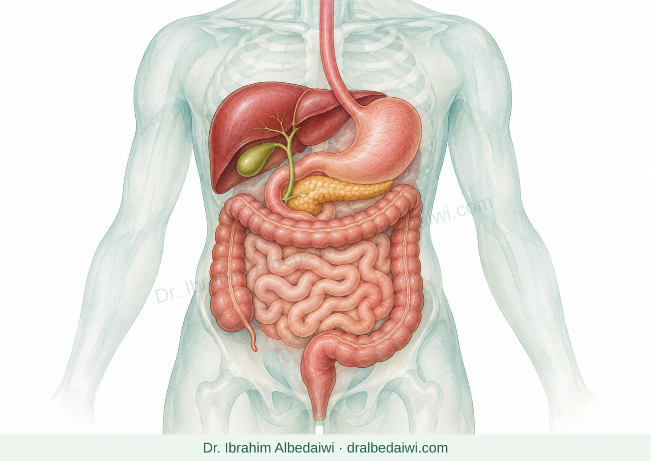

Endoscopic ultrasound
Content date: 10 November 2026
EUS combines endoscopy and ultrasound to examine the gut wall and nearby organs. It may involve imaging alone or a tissue sample; not every EUS includes a biopsy.
Your next step
Ask whether your EUS is for imaging, sampling or treatment, and confirm your medicine and results plan.
Get urgent help: Severe or increasing chest or abdominal pain, fever or chills, persistent vomiting, vomiting blood or black tarry stools, severe new swallowing difficulty, breathlessness or fainting after EUS need urgent assessment.
How is it different from gastroscopy?
- Gastroscopy shows the digestive tract lining. An EUS scope also carries an ultrasound probe, showing wall layers and nearby structures from close range. Upper EUS passes through the mouth and can examine the pancreas, bile ducts and surrounding tissues. Lower EUS examines the rectal area and has different preparation.
- EUS may clarify a small stone, cyst or mass seen on another scan, identify the origin of a lump within the gut wall, or assess the extent of a known cancer. Being referred for EUS or a biopsy does not mean you have cancer. Other scans may still be needed.
ILLUSTRATED PATIENT GUIDE
How does endoscopic ultrasound (EUS) work?
This drawing shows upper EUS.

- 1 · Scope and ultrasound probe
Enter through the mouth into the stomach or duodenum.
- 2 · Sound waves
Show the gut-wall layers and nearby organs; these are not X-rays.
- 3 · Pancreas and nearby structures
The pancreas, bile ducts and other structures can be assessed. A needle sample may be taken through the scope when needed.
Lower EUS is also used for selected problems. Not every examination needs a needle sample.
© 2026 Dr. Ibrahim Albedaiwi · Original patient illustrationsImaging or a biopsy?
- Diagnostic EUS uses sound waves rather than X-rays. If sampling would help guide treatment, the doctor may pass a fine needle through the scope to obtain cells or a small piece of tissue for laboratory examination.
- FNA means fine-needle aspiration of cells; FNB means fine-needle biopsy of tissue. The team selects the appropriate method. Not every cyst or mass needs sampling. Results are considered alongside imaging and other tests; an inadequate sample may need repeating.
Preparing safely
- Confirm whether your EUS is upper or lower, and whether sampling or treatment is planned. Follow the unit’s fasting instructions. Use bowel preparation only if specifically requested; upper EUS does not automatically require colon cleansing.
- Provide your medicine list, particularly blood thinners and diabetes or weight-management medicines. Report allergies, possible pregnancy, heart, lung or kidney disease, swallowing difficulty and previous surgery. Obtain a written medicine plan; do not stop treatment yourself. Bring previous scan and biopsy reports and arrange an escort if sedation is planned.
Can EUS also provide treatment?
- Specialist teams may use EUS to guide drainage of a fluid collection or blocked duct. This differs from imaging or simple sampling and requires its own discussion of benefits, risks and follow-up. An EUS booking does not mean you will need these treatments.
Possible complications
- Diagnostic EUS is generally low risk. Complications include sedation reactions, aspiration of stomach contents, bleeding or perforation. Sampling can add infection or bleeding risk, and rarely pancreatitis when the pancreas is sampled. Therapeutic procedures have different risks; ask about your planned procedure.
Recovery and results
- A mild sore throat or brief bloating can occur. Follow instructions for eating, drinking and restarting medicines. After sedation, avoid driving, machinery and important decisions for 24 hours, or longer if advised, and leave with an escort.
- Ask what the images showed, whether a sample was taken, when results are expected and who will contact you. An initial impression is not the final laboratory result. After treatment or stent insertion, obtain written follow-up and removal or replacement instructions.
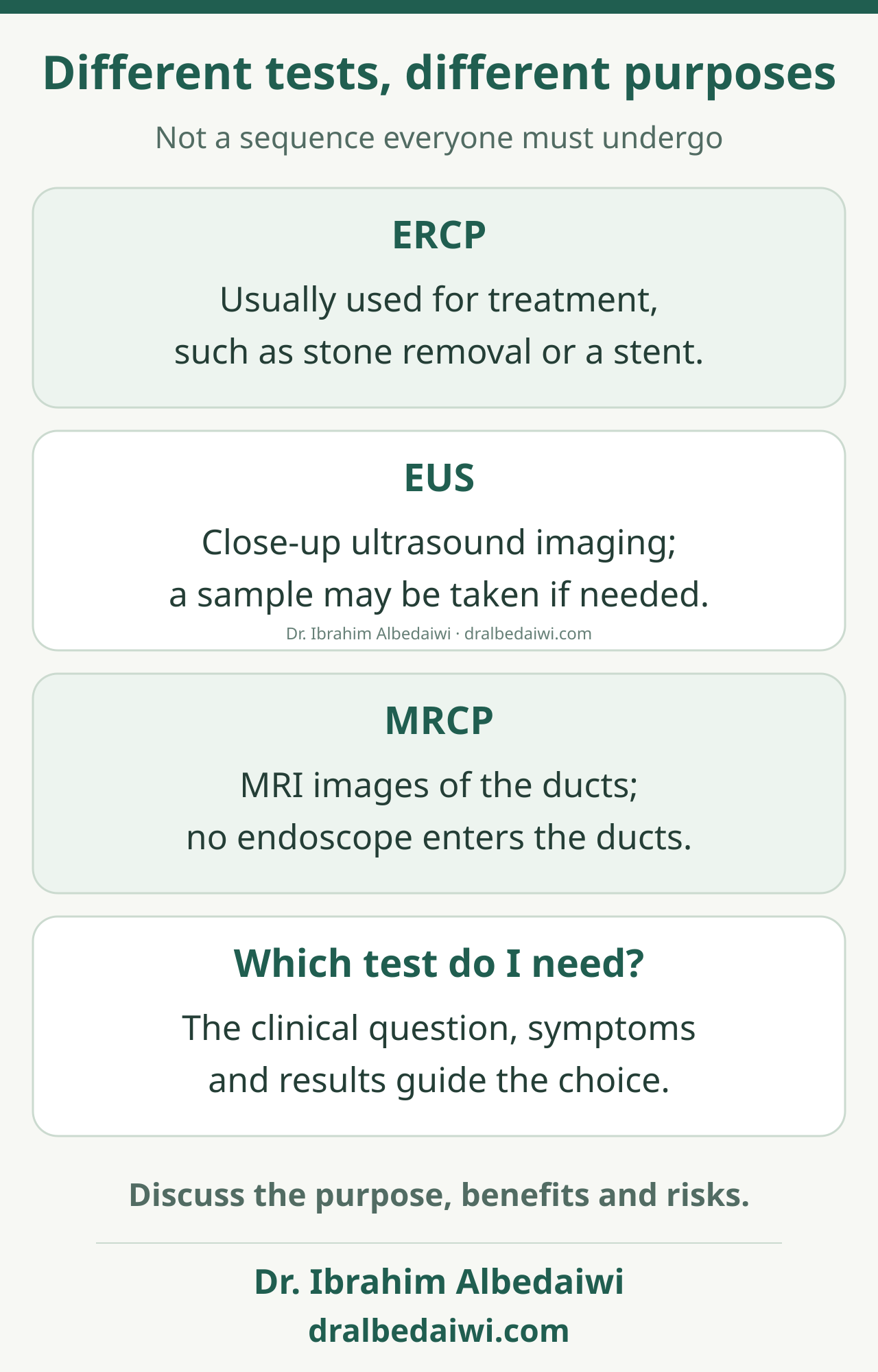
Different tests, different purposes
Read the illustration as text
Not a sequence everyone must undergo
- ERCP: Usually used for treatment, such as stone removal or a stent.
- EUS: Close-up ultrasound imaging; a sample may be taken if needed.
- MRCP: MRI images of the ducts; no endoscope enters the ducts.
- Which test do I need?: The clinical question, symptoms and results guide the choice.
Discuss the purpose, benefits and risks.
When to get help
Severe or increasing chest or abdominal pain, fever or chills, persistent vomiting, vomiting blood or black tarry stools, severe new swallowing difficulty, breathlessness or fainting after EUS need urgent assessment.
Discomfort that persists instead of improving, a missing sample result or an unclear follow-up plan. No telephone call does not necessarily mean a normal result.
Related guides
Bile and pancreatic duct endoscopy (ERCP)
Bile duct stones and obstructive jaundice